Category: Biology

  • The Hallmarks of Cancer: Growth Factors and Cell Signaling

    The Hallmarks of Cancer: Growth Factors and Cell Signaling

    This article originally appeared on Know The Cosmos. I will be re-posting excerpts here for Australian Science with added commentary over the coming weeks!

    “The Hallmarks of Cancer” are ten anti-cancer defense mechanisms that are hardwired into our cells, that must be breached by a cell on the path towards cancer. The First Hallmark of Cancer is defined as “Self-Sufficiency in Growth Signals”. What does this mean? In this post I will give an overview of growth factors and how they arhow growth signals are intimately involved in the development of cancer, it is necessary to define and understand what growth factors are, and explain how they control normal cellular behavior.

    Growth Factors
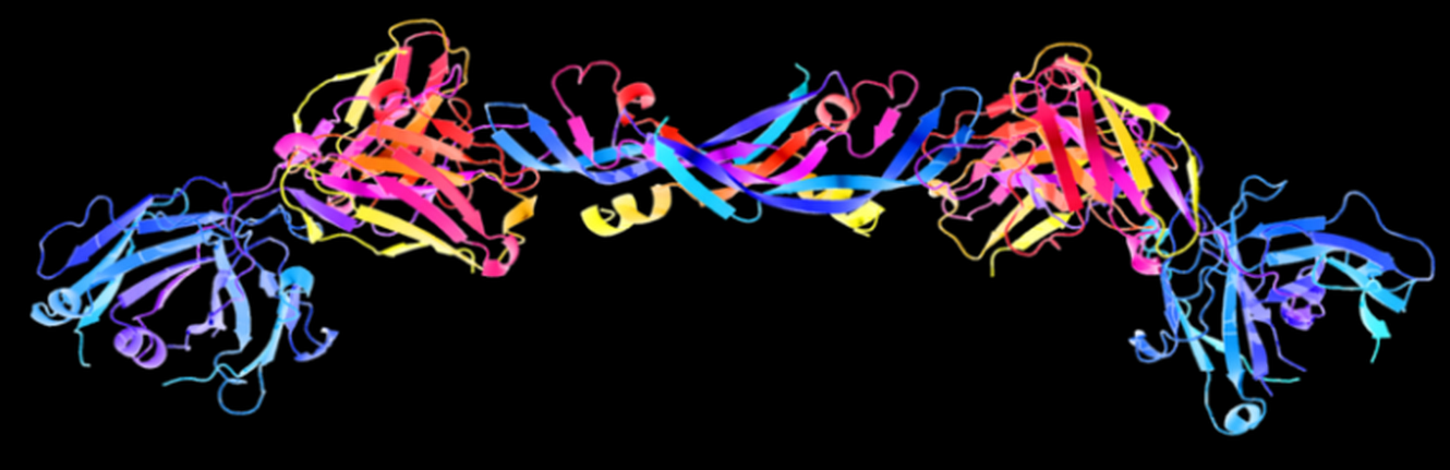
    Growth Factors are beautiful! This is a 3D schematic representation (also known as a ribbon diagram) of the structure of a growth factor known as Vascular Endothelial Growth Factor (VEGF). VEGF stimulates the development of new blood vessels, a process known as angiogenesis. Many large tumors secrete their own supply of VEGF in order to generate a supply line of oxygen-rich blood for the growing tumor to feed on. Image credit: Gizmag
    Growth Factors are beautiful! This is a 3D schematic representation (also known as a ribbon diagram) of the structure of a growth factor known as Vascular Endothelial Growth Factor (VEGF). VEGF stimulates the development of new blood vessels, a process known as angiogenesis. Many large tumors secrete their own supply of VEGF in order to generate a supply line of oxygen-rich blood for the growing tumor to feed on. Image credit: Gizmag

    Growth factors are, simply put, substances that control the multiplication of cells. There are many different types of growth factors, but they all have several characteristics in common. They are all proteins, and present at very low concentrations in tissues but with a high biological activity. They are responsible for controlling essential functions within the cell; growth, specialization and survival. Growth factors also do not circulate in the blood stream; instead, they act locally in areas near the cells that produce them. The image on the right shows a growth factor known as Vascular Endothelial Growth Factor (VEGF).

    Cell Signaling
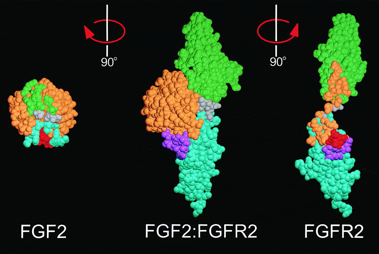
    Growth Factors fit perfectly into Growth Factor Receptor Binding Sites. Two different types of Fibroblast Growth Factor (FGF1 and FGF2, left) shown bound to its specific receptor (center) and separate (right). Image credit: Alexander Plotnikov.
    Growth Factors fit perfectly into Growth Factor Receptor Binding Sites. Two different types of Fibroblast Growth Factor (FGF1 and FGF2, left) shown bound to its specific receptor (center) and separate (right). Image credit: Alexander Plotnikov.

    It is impossible to talk about growth factors and cancer without going over some of the basics of cell signaling. We are multi-cellular animals, and as such, our cells need to communicate with each other, so they can act in a coordinated manner in response to the environment. The basis of this communication comes from a process known as cell signaling.

    The behavior of a cell depends on its immediate surrounding environment, known as the microenvironment. The assortment of growth factors in this microenvironment is the most important aspect regulating the behavior of that cell. All growth factors exert their effects by binding to a receptor. Receptors are proteins found on the surface of a cell that receive such chemical signals from the outside of the cell. Each growth factor has it’s own receptor; think of it as a key (the growth factor) fitting into a lock (the receptor). Growth factor receptors tend to be ‘transmembrane molecules’; this means that one end of the receptor ‘sticks out’ through the cell membrane into the microenvironment while the other end projects inside the cell. By spanning across the cell membrane, growth factor receptors are able to communicate signals from outside the cell (e.g. presence of growth factors in the microenvironment) to the inside of the cell. Revisiting the lock and key analogy, think of it as a key that fits into a lock that protrudes through the door-frame, instead of being flush against the door.

    The binding of the growth factor to its specific receptor triggers a phosphorylation reaction inside the cell. Phosphorylation, or the addition of a phosphate group to a protein molecule, is an important step in cell signaling. This is because many proteins exist in an ‘on’ or ‘off’ state that can be switched by phosphorylation. Therefore, phosphorylation is a key step in regulating their activity. The enzymes that add phosphate groups to proteins are known as kinases; enzymes that remove phosphates are known as phosphatases. The exterior end of the receptor protein (the bit that sticks out of the cell) carries the growth factor binding site; the other end which projects inside the cell carries a kinase site. Binding of growth factor to the receptor binding site activates the kinase domain on the interior end of the receptor protein. This activated kinase, true to it’s name, then goes on to add phosphate groups to other proteins inside the cell, which then activate more proteins downstream, triggering a signaling cascade that finally ends with the activation of genes that bring about….you guessed it, cellular growth, specialization, or survival! The image below illustrates this process – I couldn’t find a decent one online so I made my own!
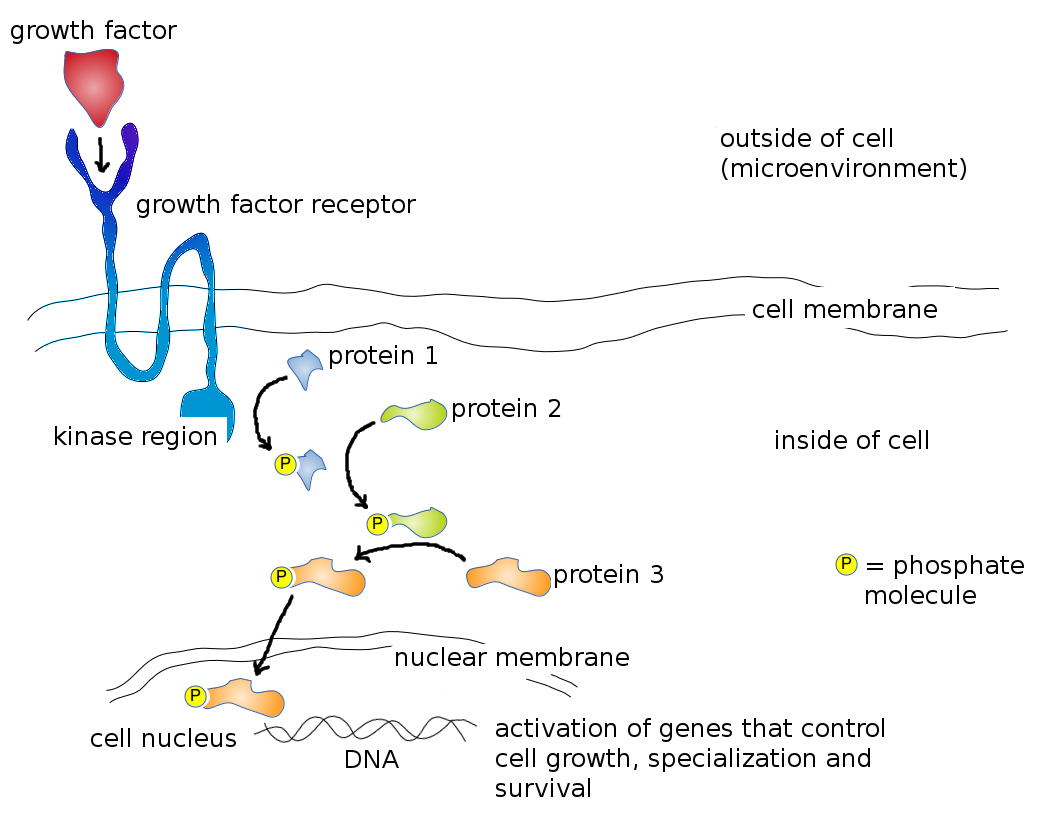
    Mode of action of a typical Growth Factor. Growth Factor (red) binds to specific Growth Factor Receptor Binding Site (dark blue) on cell surface, which activates the kinase region (light blue). Activated kinase region now adds a phosphate group (yellow) to Protein 1 (blue) which activates it. Activated Protein 1 now adds a phosphate group to Protein 2 (green) further down the pathway, which activates it. Activated Protein 2 subsequently adds a phosphate group to Protein 3 (orange) which activates it. Activated Protein 3 moves through the nuclear membrane into the cell nucleus where it physically binds to the DNA and activates genes that control cell growth, specialization and survival. Image credit: Buddhini Samarasinghe
    Mode of action of a typical Growth Factor. Growth Factor (red) binds to specific Growth Factor Receptor Binding Site (dark blue) on cell surface, which activates the kinase region (light blue). Activated kinase region now adds a phosphate group (yellow) to Protein 1 (blue) which activates it. Activated Protein 1 now adds a phosphate group to Protein 2 (green) further down the pathway, which activates it. Activated Protein 2 subsequently adds a phosphate group to Protein 3 (orange) which activates it. Activated Protein 3 moves through the nuclear membrane into the cell nucleus where it physically binds to the DNA and activates genes that control cell growth, specialization and survival. Image credit: Buddhini Samarasinghe

    The description above is an extremely simplified version of what happens inside a cell; in reality, it is not so much a linear signaling pathway as it is an interwoven, intricate signaling web, with promiscuous proteins from many different pathways activating and repressing one another. The image below is not meant to frighten you (!) but rather to give you an idea how truly complex just one such signaling pathway, known as the MAPK/Erk pathway is.
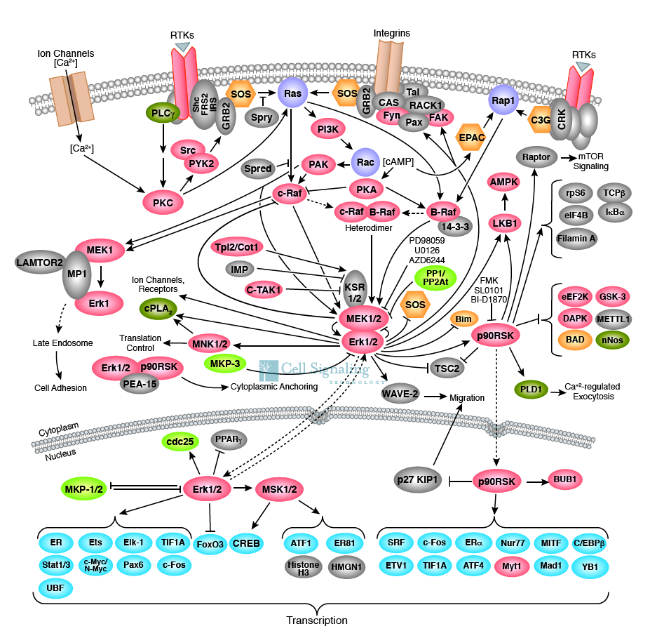
    A truly complex web of cell communication! These are some of the proteins we know that are involved in a single pathway known as the MAPK/Erk pathway. Signals from the outside of the cell go through this web of signaling, ultimately ending up with the activation of genes involved in growth, specialization and survival of the cell. Image credit: Cell Signaling Technology.
    A truly complex web of cell communication! These are some of the proteins we know that are involved in a single pathway known as the MAPK/Erk pathway. Signals from the outside of the cell go through this web of signaling, ultimately ending up with the activation of genes involved in growth, specialization and survival of the cell. Image credit: Cell Signaling Technology.

    So there you have it. We’ve covered the basics of cell signaling and the molecular mechanisms that cause a cell to grow. Next time…I will explain what goes wrong with these processes in a cancer cell.

  • The Ten Hallmarks of Cancer

    The Ten Hallmarks of Cancer

    This series of articles originally appeared on Know The Cosmos. I will be reposting the articles here for AusSci with added commentary over the coming weeks!

    In 2002, Robert Weinberg and Douglas Hanahan published a review article in the journal Cell titled “The Hallmarks of Cancer

  • Red bacteria as astronaut food

    Red bacteria as astronaut food

    Like something out of Stanley Kubrick’s famed 2001: A Space Odyssey her name is Melissa. Melissa was the first of her kind. Melissa will be there when the first of us touches down on Mars. She will feed and nurture us as we begin our exploration towards the stars. MELiSSA — or Micro-Ecological Life Support System Alternative — is a bioregenerative life-support system designed by the European Space Agency. She uses microorganisms living in interconnected controllable bioreactors to recycle organic waste. Melissa is essentially an artificial ecosystem — one that turns waste into food to feed her crew.

    When long space missions start to become commonplace, when we start colonies over the horizon, one of the obstacles will be how to be self-sustaining in space. Within a closed arti

  • The language of biofilms

    The language of biofilms

    Every once in awhile, communities form. Collections of similar or diverse things come together, and in unison strive for a common goal. It is the same for mice, men, and bacteria. Humans do it and build nations. Bacteria do it and build biofilms.

    Dental plaque, the slimy coating on pipes and tanks, algal mats on a still lake — are all different types of biofilms. Biofilms for us are a nuisance because they colonise medical devices implanted in the human body.  They can be used, however, in treating sewage, industrial waste, or contaminated soil.

    When a biofilm forms, it is far from random. At its leading edge — the biofilm has purpose and direction. Imagine it as an invading army sending out the vanguards toward an unexplored territory. Highly coherent groups of bacteria migrate across the surface — swarming as one. As they advance, they create furrows for those bacteria at the back to follow. The vanguards carve out a network of trails — one that will eventually guide the exodus — the mass transit of following bacteria towards the leading edges of the biofilm.

    Pseudomonas aeruginosa — the bacteria they use to eat up oil spills — is able to colonise many natural and artificial environments. It thrives on most surfaces, and easily causes a problem for implanted medical equipment like catheters. Individual bacteria show distinctive multicellular behaviour. When they grow, patterns and order emerge from seeming chaos. Australian scientists, publishing in the Proceedings of the National Academy of Sciences, describe the methods they used to visualise movements of individual bacteria, and to characterise the order within. Researchers had to develop sophisticated computer algorithms to visualise, identify and track individual bacteria. Carrying out a time lapse recording of bacteria at one frame every 2 seconds, and visually inspected a 1000-frame time series (download the movie).

    As the bio

  • What Animals Will Go Extinct During Our Lifetime?

    What Animals Will Go Extinct During Our Lifetime?

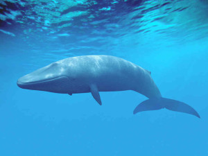
    Blue Whale, via NPR.org
    Blue Whale, via NPR.org

    I spent the other day meandering through some of the exhibits on display at the American Museum of Natural History. I checked out the Global Kitchen, Butterflies, and Whales exhibits and the planetarium Journey to the Stars show. Between the whales and the planetarium, I had this thought: What animals will we see extinct during our lifetime?

    Perhaps a morbid thought, but when you walk through an exhibit on whale hunting, see hundreds of taxidermic specimens, stroll through the hall of biodiversity, you start to think about the state of the natural world. And what a state it is in.

    Whales

    The sheer size of a blue whale (30 metres) or a sperm whale (15 metres) is astonishing. Several skeletons were on display at the museum. A model of the sperm whale’s heart – about the size of a Mini Cooper – serves as an indication as to what these giants of the sea need to survive from an anatomical and physiological perspective. What they don’t need to survive was evident in photos showing the flesh of whales sliced through from the blades of a shipping container’s propeller. The mysterious mass beach strandings, fishing nets, oil spills, the Japanese “Research

  • The parasitic warfare perpetrated by ladybirds

    The parasitic warfare perpetrated by ladybirds

    There is a scientific term that causes fear and alarm to those that study biodiversity. More fear and alarm than the term climate change. Biotic homogenisation — introducing a new exotic species to an area that was, until now, without admixture. Worldwide there is an increase in introduced exotic species and the potential of these species to become invasive have their impact.

    The most invasive ladybird on Earth — Harmonia axyridis — was introduced in several European countries and North America for biological pest control, and quickly turned invasive. It has been outcompeting indigenous ladybird species in many countries for a long time now. Ladybirds are quite common in use as a — what we now call — biological control agent. They have a long history of use against unwanted pest insects. For example, the Australian vedalia ladybird, Rodolia cardinalis, was released in 1888 to control scale insects.

    Harmonia axyridis is also known as the harlequin ladybird, and it has been outcompeting and threatening native species since the beginning of the 20th century. Its invasive success has until now been attributed to its enduring resistance against diverse pathogens. The same pathogens that strike down and blight the native European species, allowing the harlequin ladybird to outperform and out-survive.

    But now, Andreas Vilcinskas knows of another reason — and it is something more like parasitic warfare than simple evolutionary survival of the fittest.

    Andreas Vilcinskas goes to work every day to kill ladybirds. Or rather, he goes to work to investigate how ladybirds kill each other. And it is the parasites within that act as the smart bomb against the native species. Harmonia have within them, swimming around in their blood, spores of the parasite microsporidia. They do no harm to Harmonia, but are lethal to the native ladybird Coccinella septempunctata. What is more interesting is that this lethality comes when the native species feed on microsporidia-infested Harmonia eggs or larvae. As is convention, ladybirds often eat the eggs of other ladybirds.

    The tale of an invasive species is always something more akin to Kal-El leaving Krypton. You land in a foreign land with the realisation that you have superpowers (in comparison to the natives). The harlequin ladybird’s secret most likely lies within its blood (hemolymph). A hemolymph already shown to ward of mycobacterium and the parasite that causes malaria. Its blood, or more accurately, the metabolite harmonine within its blood, exhibits broad-spectrum antimicrobial activity. Antibacterial activity against Mycobacterium tuberculosis, and chloroquine-resistant Plasmodium falciparum strains.

    For this new reason to awe the ladybird, Andrea Vilcinskas and colleagues drew blood from the legs of 100 ladybirds. With such super blood already documented, the question being: was the harlequin ladybird’s advantage over the native species simply down to harmonine, or did the microsporidia have a significant lethal effect.

    They showed that injecting the native species with Harmonia blood — but not harmonine alone — can kill. Suggesting, that it is unlikely that the mortality caused by native species feeding on Harmonia eggs is caused by the presence of harmonine.

    It seems that native ladybird species are lethally infected with microsporidia carried by Harmonia when they feed on its eggs and larvae. The microsporidia parasitize cells by means of a spear through the plasma membrane. Within two weeks of starting their experiment all native species had died from the microsporidia.

    The most invasive ladybird on Earth lives up to its name. The worldwide invasion of the harlequin ladybird is as a result of a parasite that lies within. A parasite to which it has grown immune but to which the natives have no way of stopping. War of the Worlds gone backwards.

    Image — source.

  • The beat the mosquito’s heart didn’t skip

    The beat the mosquito’s heart didn’t skip

    It pulsed continuously without stopping. Then it repeated, as it had done many times before. Then, without delay, almost without skipping a beat, it changed direction. The action was as old as man himself, yet this time, completely and uniquely different — the perfect heartbeat. The perfect mosquito heartbeat.

    An interesting quirk of nature is how remarkably constant the number of heartbeats exist within a lifetime. An interesting quirk, more a function of the metabolic demands of the animal in question rather than any underlying feature of the heart itself. Humans, mice, insects all have the same number of total heartbeats. The human heart, from life to death, will beat roughly 3 billion times. A mouse will use up its heartbeats in about two years. An elephant, with a much slower heartbeat, will last for much longer. The mosquito’s heart beats at a rate of just over one beat every second (1.3 Hz). In one minute it will beat 82 times, of which, some of that will be in the other direction.

    The heartbeat is nothing unique to humans and has been around long before us, but we have romanticised it and given it a meaning more than its basic function. For researchers at Vanderbilt University, Nashville, Tennesee, this is also the case for the mosquito’s heart, where function and meaning is more than its basic, simple architecture.

    Dr Julian Hillyer, the lab’s director, and his team have offered the most comprehensive visualisation to date of how the mosquito’s heart beats. They filmed live restrained female Anopholese mosquitoes — the same species of mosquito responsible for life threatening malaria — through a microscope connected to a very sophisticated camera.

    The beatings of thirty mosquitoes were collected and analysed frame-by-frame to arrive at a comprehensive structure of the heart. They painstakingly dissected individual mosquitoes, injecting infinitesimally small amounts of fluorescent fluid into the mosquito, allowing them to describe the mechanics, directionality and flow involved when the insects blood (hemolymph) is propelled through the heart.

    A mosquito’s heart is very different — without veins or arteries, it pumps a clear liquid called hemolymph. The hemolymph flows from the heart into the abdominal cavity and eventually cycles back through the heart. The heart runs along the insects body as an unbranched tube, no thicker than three tenths of a millimeter. Helical twists of muscle fibres support the central tube. Their sequential contractions makes the heart in a wave-like peristaltic action. A peristaltic action that has the ability to run in both directions.

    Another set of muscles anchors the heart where ever there is a valve, at intervals, along the mosquito’s body – just underneath its cuticle shell. All of this was visualised in fluorescent detail, using different coloured flourescent dyes to highlight different structures inside the insect’s body. Winning the lab’s images the Nikon Small World photomicrography competition in 2010.

    As stunning as the images were, it was the functionality gained from the study that provided the most insight. Following and tracking tiny microscopic particles (microspheres) showed how the insect’s hemolymph entered and was expelled from the heart, and, most importantly, how the heart reverses direction.

    Most of the time, the heart pumps the mosquito’s clear hemolymph blood towards the mosquito’s head, but occasionally it reverses direction and pump fluid to the last segment of its abdomen. The direction in which the heart contracts reverses roughly 5 times every minute.

    Heartbeat reversal is not unique to the mosquito — a phenomenon that has been observed in other orders of insects. You would think that something that small would have no need for such an elaborate beating system, but perhaps it is the only way the heart can regulate the different hemolymph pressure and volumes entering it. Thus far, a conclusive “why

  • Combating the rise of the superbugs: The health and scientific challenges of antibiotic resistance

    Combating the rise of the superbugs: The health and scientific challenges of antibiotic resistance

    It’s hard to imagine the world prior to antibiotics, a world where even a deep laceration could frequently spell significant illness or even death due to infection. Thankfully, since the discovery of penicillin in 1929 by Alexander Fleming, we now have a range of potent antibiotics to treat many of the various types of bacterial infection.

    There is a problem though, bacteria are great survivors and have been competing against other bacteria and microorganisms for billions of years. As  Professor Matt Cooper from the University of Queensland  puts it “Billions of years ago, bacterial species were engaged in an arms race against each other and the chemicals they developed to kill one another have been modified into today’s antibiotics”.1

    Multi-Drug resistant Tuberculosis is a particular concern for health authorities and clinicians due to limited treatment options.

    Unfortunately it’s our overuse of these important drugs which has driven the rapid development of antibiotic resistance, the process whereby bacteria containing mutations in their DNA, that provide some protection from an  antibiotic, have an enormous survival advantage when exposed to the antibiotic and pretty soon dominate. Frequent exposures to the antibiotic may further strengthen these survival traits via the selection process, rendering the the drug less effective over time. It’s a great example of random variations leading to non-random adaptions through natural selection, although one with profound consequences for human health.

    Of particular concern are bacteria that have developed resistance to multiple types of antibiotics, resulting in particularly dangerous resistant bacteria such as the multi-drug resistant variants of tuberculosis, that are extremely difficult to treat. Indeed leading health authorities are so worried about the problem that the Chief Medical Officer of the UK, Professor Dame Sally Davies, has recently labelled the threat as “catastrophic

  • Invasion of the Asian tiger mosquito

    Invasion of the Asian tiger mosquito

    Sometime during that glorious decade known as the 1980s, a shipment landed in Houston, Texas. A shipment carrying more than its cargo. The point of origin was Japan. The shipment was used tires. The payload was Asian tiger mosquitoes.

    Within years of landing in Texas the tiger mosquito, Aedes albopictus, rapidly displaced resident populations of Aedes aegypti mosquito. Both are important disease vectors. The native being considered the primary vector of breakbone fever — or dengue. And the Asian tiger mosquito recently emerging as the most important transmitter of chikungunya virus and yellow fever. By September 1986, the range of the Asian tiger mosquito had extended as far north as Utah. The demise of the native Aedes aegypti had already begun — representing not a transient ecological phenomenon, but the beginning of permanent colonisation, and resulting in rapid declines and extinctions of the native mosquito species.

    Aedes aegypti originated in Africa and was introduced to the Americas between the 15th and 18th centuries, during the height of the slave trade and most likely on ships transporting slaves. They quickly established across the south eastern part of the US. Then what followed was a demise in the face of stiff competition and after centuries of habitation in America. An inadvertent metaphor for the “Post-America

  • Rare echidna species not so extinct after all?

    Rare echidna species not so extinct after all?

    Speaking as a European, Australia has something of a reputation for having some rather unusual wildlife. Easily the most unusual are the small handful of monotreme species – the echidnas, and the duck-billed platypus. The only species of egg-laying mammals in the world today, these little creatures may once have been quite widespread. Now, however, they’re only found in Australia and New Guinea. One species in particular, the long-beaked echidna, is critically endangered. It was believed to have been extinct in Australia for over 30,000 years (and only found in New Guinea), since the last ice age. It was believed, that is, until recently. And the evidence for this rediscovery came from a rather surprising source.

    100 years ago, biologists worked a lot differently to the way they do today. Back then, it was common practice to travel to remote places and collect specimens – by way of hunting animals, shooting them, and getting a taxidermist to stuff them. While this bloodthirsty pokemon attitude may seem ghastly to our modern sensibilities, it was once simply the way things were done, and many such specimens are still on display in museums. Though it should be added that such specimen collecting is widely outlawed today.

    Nonetheless, one such specimen was found in London’s Natural History Museum. The creature had been “collected” in Australia in 1901, scientifically described, and had subsequently been stored and forgotten about entirely. I have to wonder what those researchers may have done if they’d realised the true significance of this unassuming little creature.

    The fascinating thing is really that this little preserved creature is the keystone for the entire study. Just one single specimen. However, it was very well documented and most certainly came from Australia. Its discovery was quite serendipitous too, when zoologist Kristofer Helgen from the Smithsonian Institution, Washington, was paying a visit to the London Natural History Museum.

    From the description it was tagged with, this echidna had been found on Mount Anderson, in sparsely populated Northwest Australia. Following up the find, researchers decided to investigate further. In West Kimberley, they spoke to some aboriginal communities where people recounted stories of how their parents used to hunt echidnas which were much larger than the others. Using photographs, they identified those large echidnas as the same long-beaked echidna species still found in New Guinea.

    So the big question is, are long-beaked echidnas still found in Australia today? This discovery does give us some more information about how adaptable these spiny little animals are; long-beaked echidnas can evidently survive in both arid Australian scrub land and lush New Guinea rainforests. Until a living animal is found, it’s impossible to make any definite statements. And finding them is no easy task. They’re nocturnal creatures, and the known populations of them in New Guinea are difficult to find. All the same, conservationists can be hopeful that long-beaked echidnas may not be extinct in Australia just yet.

  • The bacteria that live inside hurricanes

    The bacteria that live inside hurricanes

    Seven miles above the Earth’s surface, where the weather is born, lies the troposphere – the lowest layer of Earth’s atmosphere. Up there, where the clouds dance around, are bacteria that can make it rain, and are important for the formation of clouds.

    The atmospheric microbiome is a concept and field of study that is gaining importance. As we come to grips with a changing climate and environment, understanding more and more our Earth ecosystem remains vital. With hurricane damage in the US and elsewhere seemingly on an exponential increase in recent decades, it is important to mitigate for the worst. It can cost as much as $1 million per square mile for evacuation preparations alone.

    In 2010, NASA embarked on one of its largest hurricane research efforts — GRIP (Genesis and Rapid Intensification Processes). The objective was to better understand and characterise how tropical storms form and develop into major hurricanes. With a fleet of aircraft, ground-based instruments, computer models, and satellites, over a period of 6 months, GRIP collected all kinds of data on the nature, structure, dynamics, and motion of hurricanes. Invaluable data. They also collected one other thing — the microorganisms in the atmosphere.

    The problem previously, had always been the difficulty in gathering enough microbial biomass to study. And previously, most samples have comes from areas too close to the Earth’s surface to really mean anything. GRIP took things one step further — high-altitude. Over the course of 9 flights across America, the Gulf of Mexico, the Atlantic Ocean, and the Caribbean, GRIP collected bacterial and fungal samples to be analysed. Enough to answer the question: Where does the bacteria in the atmosphere come from? Authors, publishing in the Proceedings of the National Academy of Sciences (PNAS) today give a picture of the composition of the high-altitude (around 10 kilometres above sea level) bacterial and fungal flora, but also what that picture looks like in the aftermath of a hurricane.

    The bacteria that swirl around in the air originate from different areas across the Earth’s surface it seems. The organisms they sampled originated from almost all habitats (ocean, soil, freshwater… etc as they put it). Hurricane samples had a higher abundance of marine bacteria, and only in the hurricane samples was there “a substantial signal of bacteria known to be associated with human and animal feces

  • The animal link to sleeping sickness

    The animal link to sleeping sickness

    As with many parasites, the nuisance they bring is partly compensated for by new insights they provoke. The African trypanosome is perhaps unique among all of the diseases of developing worlds. The diseases of sleeping sickness, inflicted on man and cattle alike, perhaps drove early man ‘out of Africa’ — in an attempt to avoid tsetse infested areas of the Rift Valley. The Zulu word for powerlessness and useless, “N’gana

  • Citizen Science

    Citizen Science

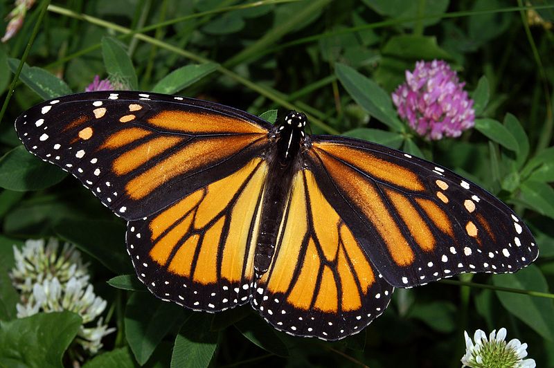
    Monarch butterfly (Danaus plexippus), Source: Wikipedia Commons

    Citizen Scientists

    An interesting report released last week from the UK Environmental Observation Framework reveals the benefit of citizen scientists to governmental environmental organisations. The Nerc Centre for Ecology and Hydrology and the Natural History Museum in London, reviewed 234 projects – ranging from small surveys to large-scale programmes. The results found the involvement of volunteers offers “high value to research, policy and practice”.

    Given the increasing number of complex scientific problems facing our communities, states and countries; coupled with the ever dwindling supply of cash on hand to fund research projects, it behooves governments to dial into this untapped source of volunteer environmental monitors and data collection specialists. People enjoy going to their local botanical gardens, taking the kids and grandkids to learn about the natural world surrounding them. A large majority of the population love their parks for taking walks and having picnics and for exercise. They want to see these lands, with the flora and fauna contained within them protected. People volunteer for things they believe in and causes they want to see sustained. That is the movement around science and the environment and why the citizen scientist movement is growing.

    Scientists should be working with their governmental departments, or university outreach extension centers, to coordinate community science volunteer days. Measuring leaf litter in a forest, or tree ring growth, or monitoring a stream for the presence of certain harmful bacteria or toxic chemicals, or tallying the number of a threatened bird species are a few such projects that could be undertaken by community members. Scientists and their research teams and graduate assistants could customize their research projects with a component where they served as project managers working with the community to accomplish a piece of the data collection puzzle.

    The Value – Minuses and Pluses 

    Of course, not every project will be appropriate for citizen scientists and a fair amount of training may have to be done, given the nature of the project. And the report touches on the fact that although the quality of the data collected by citizen scientists could be excellent, it may not be fully recognized by all researchers or policymakers. This seems to be an area that would require some policy work in setting up standards to ensure the legitimacy and recognition of such projects undertaken with contributions from citizen scientists. That minus shouldn’t be so hard to turn into a plus. Volunteer-collected data possesses a bevy of potential benefits. The use of smartphones continues to increase among consumers and with that comes development technologies in the form of apps that could play an easier role in data collection. A scientist could quite easily dispatch his volunteer army with marching orders to perform observations of the monarch butterfly (Danaus plexippus) over a 5-acre area of the park and have his/her data collection completed within a month’s time. And of course, the benefit that every bureaucrat or department chair loves to hear: it’s a cost-effective way to collect environmental data and could help meet the demands of increasing governmental reporting and compliance requirements.

    Getting Started

    I think it’s up to each and every one of us to identify the actions we can take that will help continue to make our environment habitable. And becoming involved in a citizen scientist program seems like an ideal way to start to make a difference in your community. Whether you are a scientist or a volunteer, the guide, produced alongside the report by the UK Environmental Observation Framework, is a great place to begin identifying and mapping out strategies for a collaborative citizen science project.

  • Does my science look big in this? The astrobiology edition

    Does my science look big in this? The astrobiology edition

    During the 20th century a powerful new idea gradually entered our consciousness and culture: cosmic evolution.  We are all par of a huge narrative: a cosmos billions of years old and billions of light years in extent. It is this idea that caught my attention this month via the proceedings of the Sao Paulo Advanced School of Astrobiology SPASA 2011, published in the October International Journal of Astrobiology.

    Although the question of extraterrestrial life is very old, the concept of full-blown cosmic evolution – the connected evolution of planets, stars galaxies and life on Earth and beyond – is much younger. In a rather breathtaking paper, Steven Dick formerly of the Aerospace History at the National Air & Space Museum places his arguments for cosmic evolution. Dick traces the idea from its roots in the 19th century theories of Pierre-Simon Laplace and Robert Chambers through its philosophical, astronomical, and biological upbringing to the present day. He examines evolution, the worldview that it had become in the 1950s and 1960s and how it had permeated culture in numerous ways and different cultures in diverse ways. Dick cautions us though noting “we need to remember that ‘culture’ is not monolithic and that ‘impact’ is a notoriously vague term.”

    Cosmic evolution. Image credit: Harvard University.

    In addition to the impact of our new understanding on culture, cosmic evolution also provides a window on long-term human destiny, asserts Dick. He presents this idea via three scenarios, the: the physical , biological, and postbiological universe. Life is unique to earth in the physical universe scenario, and the options flow from this situation – think of Isaac Asimov’s Foundation series. We will certainly interact with extraterrestrials in the biological universe – here cosmic evolution commonly ends in life, mind and intelligence. Cultural evolution in a biological universe may replace biologicals with artificial intelligence creating what Dick calls a postbiological universe. We do not know yet, which of these is our reality, that is one of the challenges of astrobiology, maintains Dick.

    In a second ‘big-picture’ paper Marcelo Gleiser presents his four ages of astrobiology. For Gleiser the influx of astrophysical data, particularly on the prevalence of exoplanets “indicates that there are plenty of potentially life-bearing platforms within our galaxy.” He then presents the ‘history’ of life in the universe in terms of the steps needed for matter to have sequentially self-organised into more and more complex structures. His sequence is best viewed as a prelude to the physical or biological universe scenarios of Dick. Gleiser’s fourth age, the Cognitive Age (the age of thinking biomolecules), really addresses whether we are unique or not i.e. which of Dick’s two scenarios, the physical or biological are reality. Gleiser’s first three ages: physical, the creation  of stars and planets from atomic nuclei; chemical, in which elements organise into biomolecules; and thirdly biological, in which living creatures of growing complexity form from biomolecules. the papers by Dick and Gleiser are both papers heady and exhilarating conceptual reads.

    Jorge Horvath and Douglas Galante accept the premiss that life exists, and then argue we need to take high-energy astrophysical events seriously. Scientists and the public account for meteor impacts in both academic studies, science-fiction writing and film – not so for events such as supernovae, gamma-ray bursts and flares. They show that these events are more frequent than asteroid strikes and that the effects are non-negligible (academic speak for potentially fatal to planet based species). They conclude that just because we have not yet been wiped out by such events can be seen as either a measure of earthlife’s resilience or a threat we are statistically yet to encounter.

    My attention was captured by two other papers from the proceedings. Martin Brasier and David Wacey address the problem of studying life in deep space – comparing it to study of life remote in time. This view is pertinent, as it is non-trivial for scientists to determine what is a viable signal of extinct life. The authors develop a set of protocols and then apply these to earth samples, of varying ages. They do this to show how we could interpret similar samples, where much of the desirable information (the context) has been filtered out during the process of transmission (either physical or data) across vast distances of space, or time or both (as is likely on Mars). Even 10 years ago these questions were moot, but we have learned much over the recent past about metabolic pathways and living microbial systems. Brasier and Wacey conclude that there is still work required on pseudo-fossils, structures that arise naturally within complex physico-chemical systems, so that we can confidently agree on signs of life that are remote in space and time.

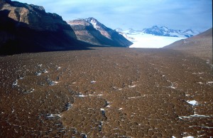
    The Dry Valleys in Antarctica. Photo credit: NASA

    My final pick is an experimental paper that looks at the ExoMars mission. The European Space Agency and (initially NASA ) ExoMars mission is scheduled for launch in 2018 – specifically to detect life signatures on the surface and subsurface of Mars. This probe will carry, for the first time, a Raman spectrometer,  a technique with proven ability to determine the spectral signals of key biochemicals. The authors support these assertions by assessing samples acquired from Arctic and Antarctic cold deserts and a meteorite crater. These terrestrial environments are similar to those found on Mars. The experimental results presented in this paper demonstrate that it will be possible using this technique to assess and detect spectral signals of extra-terrestrial (Mars in this case) extremophilic life signatures.